Basics of Digital Dental Photography
In today’s society of patient’s high expectations and increased demands for cosmetic dentistry, dental photography is essential for :
– Diagnosis and treatment planning: As a dental professional you gain a more detailed information for comprehensive treatment planning.
– Patient Communication: Patient ability to understand their needs and complications is much better when viewing their own pathology.
– Lab Communication: Technician receives a full range of shades instead of single shade. Better aesthetics and colour achievement.
– Interdisciplinary Communication: Complex treatment planning is easier between different specialities with proper documentation.
– Legal document/Record keeping: Dental photography is a way of documenting patient’s records in case of a need for legal defence.
– Clinical research/Teaching: Images can be used for future references.

In order to obtain adequate and satisfactory results, proper technique, camera, lens, lighting and accessory equipment is required.
Things You’ll Need:
1. Digital SLR camera body
2. Macro SLR lens
3. Light Source
4. Intraoral Mirrors
5. Lip and Cheek Retractors
6. Anterior Contraster (optional)
1. Digital Slr Camera Body
Single lens reflex (SLR) is a perfect camera for all-round photography. It allows control over every aspect of the picture taking process when you feel it is necessary, but also it can make all the decisions for you. The single lens refers to using one lens for capturing images and displaying them on viewfinder. The reflex part refers to the use of a reflex mirror which reflects the image passing through the lens towards the viewfinder. This mechanism allows you to see exactly what will be captured by the film or sensor without parallax or distortion. The most difficult part of photography for a beginner is actually deciding which type of camera to buy.
2. Macro SLR Lens
Macro lens is a specialized lens that allows very close focusing without the use of any close-up lenses or extension tubes. It has fixed focal length and is probably the most important part of dental photography. While purchasing a macro lens one should always remember to purchase a lens with reproduction ratio of 1:1. Since clinical photography needs a focal length of 95-110 mm, the recommended lenses are:
1. Canon EF 100 mm f/2.8 macro USM
2. AF micro Nikkor 105 mm f/2.8D 3. Sigma 105 mm f/2.8 EX DG macro

3. Light Source
If the subject is not lit adequately for proper exposure than electronic flash for illumination is needed. In dentistry, we are working in an oral cavity which is quite deep and has variety of areas casting shadows on each other. For this reason, we need light source which could work in close-up proximity and eliminate shadows. The light sources that are specifically designed for this purpose are Ring Flash or Dual-Point Flash. Both fit on the front of a lens and have a light or bulbs to provide even, shadowless illumination, as they throw light from all directions.

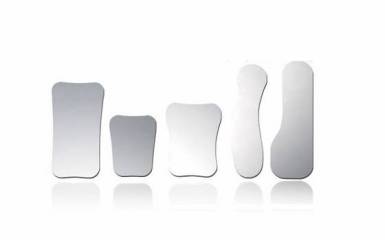
4. Intraoral Mirrors
Intraoral mirrors permit the indirect photography of areas of the mouth which are not directly accessible and, if correctly placed, alleviate the problem of depth of field. There are two types of photographic mirrors.
– Metal mirrors are less expensive, robust, and can easily be sterilized in an autoclave. Optically, they are inferior to glass mirrors, especially on the edges.
– Metal-film plated glass mirrors are more fragile and expensive, but yield far more brilliant mirror images and are therefore a preferred option. These should be cleaned and disinfected carefully to avoid damaging the delicate metal coating. 
Occlusal mirrors are used for maxillary/mandibular occlusal images.
Buccal mirrors are used for quadrant, buccal, and lingual images.
Some points in order to achieve good images using mirrors:
– The image should be framed so that only the mirror image of the teeth is captured. The image can then be reversed and resemble a photograph which was taken directly.
– Structures or edges of the mirror should be hardly or not at all visible. For that reason, you should try to use the biggest mirror possible.
– The fingers holding the mirror should be as far to the front as possible so that they do not appear in the photograph. Mirrors with handles are an advantage.
– Fogging on the mirror’s surface can be prevented if an assistant warms the mirror first or directs air at it.
– The patient should be instructed to breathe in through the mouth and out through the nose during photography.
– Finally, mirrors should never be placed on or near metal instruments to avoid scratching.

5. Lip and Cheek Retractors
Opening the oral cavity with the use of retractors is necessary in order to access the zone to be photographed and to achieve optimum illumination. The most commonly used lip holders are those made of clear plastic; these are available in a variety of size, are most comfortable for the patient and, if photographed, do not interfere with the image, since they allow the underlying structures to shine through. They also have the advantage that their size and shape can be altered. Retractors made of wire are also in use, which have a larger or smaller bend at either end. The disadvantage here is that the centre of the lips is not held and that the highly polished metal can cause reflections. Lip retractors made of metal (not wire) are not recommended, since the reflections from them cannot be controlled and spoil the image. They can also cause incorrect exposures when using flash, because the flash sensor can be “fooled” by the strong reflections.
Some points in order to achieve good images using retractors:
– Lip holders should generally be used in pairs.
– As a rule, the largest lip holder possible should be used to open the mouth as wide as possible. The size depends not only on the size of the oral cavity, but also on the tone of the lips.
– Cream should be applied to dry and cracked lips beforehand to avoid lesions.
– The lip holder is moistened, inserted over the lower lip and carefully turned into the corner of the mouth. In so doing, no pressure should be exerted on the gingiva.
– Retractors should usually be positioned by the doctor, and held by the patient.
– They should not be visible in the photo.
6. Anterior Contraster (optional)
Used in anterior shots to “black out” the background to enhance the ability to see translucency. They are used in conjunction with retractors. Finally, regardless of the equipment you chose, familiarity and practice with your camera system will eventually produce good results. At the end of the day, one must remember that camera is only as good as a person using it. Cameras only deal with the mechanical side of photography; they cannot compose pictures, choose subject or tell when the light is right. Those decisions will always have to be made by you, and they are, by far, the most important points.


Really useful article! Similar to my recent post too! Best wishes
http://atoothgerm.blogspot.co.uk/2014/10/a-guide-to-photography-in-dentistry.html
LikeLiked by 1 person
Hi Natalie,
I really enjoyed some of the posts in your blog. Thanks for the link.
Happy NEW YEAR 🙂
LikeLike
Thanks so much! You too! Good luck in 2015!!
LikeLiked by 1 person
Reblogged this on DrBruschi and commented:
Nice work from Kasia
LikeLiked by 1 person
Woah !! Enjoyed reading every word.
LikeLike